Overview advantages

- Fully automated detection
- Options to include or exclude regions or cell groups using freehand selection
- Automatically calculate scores
- Reports instantly available: Analysis reports available as image and text
Due to the high detection speed, accurate results are available after only a few seconds.
The use of slide scanners is possible, but not mandatory.
The existing microscope and a microscope camera can be used just as well.
Our system runs without internet connection and even on older systems.
- University of Applied Sciences, Berlin
- Charité University hospital Berlin
- Internationale Academy of Pathology – german divison e. V.
- Charité University hospital Berlin Centre for Anatomy
- The Physikalisch-Technische Bundesanstalt (PTB; National Metrology Institute) in Berlin
- Dr. Senckenbergisches Institut of Pathology, Frankfurt/Main
- Institute of Pathology Kantonsspital St. Gallen
- Institute of Pathology in Kantonsspital Winterthur
- Institute of Pathology Würselen/Aachen
- Düren Pathology
- Institute of Pathology
Klinikum Dortmund


Cognition Master Professional Suite
Digital Pathology System
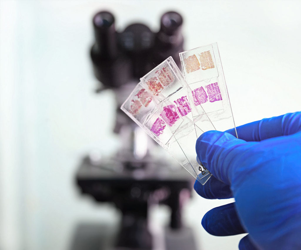
Cognition Master Professional Suite is a platform for digital pathology and image analysis. The modules have been developed in co-operation with our partners from pathology and automatically evaluate images from your microscope camera. To save time and reach highest accuracy.
Digital Slide Suite
Web Slide Portal
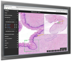
Digital Slide Suite is your solution to bring own slide collections for example for research or presentations to the internet, without own efforts. The web microscope runs on all devices: Fully capable for PC, tablet, smartphone. And without any brwoser plug-ins.
VM Slide Explorer
Digital Microscope as local application
VM Slide Explorer is a digital microscopy application that allows you to navigate virtual slides efficiently on the monitor. At the same time you can make use of the imaging modules of CognitionMaster Professional Suite. Furthermore snapshots facilitate the assembly of digital documents and presentations.VM Slide Explorer is integrated into several information systems and LIMS. For an easy and ideal workflow.

VM TMA Evaluator
TMA Evaluation on Virtual Slides
VM TMA Evaluator allows to evaluate digital Tissue Microarray (TMA) slidess. That includes the automatic detection and assignment of TMA spots on the scanned TMA slide. The evaluation follows an evaluation scheme that can be defined very flexibly. The evaluation results can be exported as MS Excel tables.
VM Slide Converter
Converting Virtual Slides
VM Slide Converter generates virtual slides in the standard format JPEG2000 or as bitmap from the file formats of several slide scanners. That allows you to use your virtual slides in several applications or to exchange it with colleagues, which might not use your software.
„Digital Slide Suite enabled us to digitally provide the entire case with specimens for the students. Every student in the lecture room sees the exact same slide including annotations. The students also enjoy the opportunity to learn about the slides from home via the Internet.“
We look forward to your inquiries and questions about our products and services.


We would be glad to answer your questions in a personal meeting or via e-mail.
No, our solutions run local. (Depending on the features you want to use, you need a connection)
Good news: our solutions are designed to work also on standard pc systems.
The use of slide scanners is possible, but not mandatory.
It is just as well to work with the existing microscope and a microscope camera.


